Retinoscope
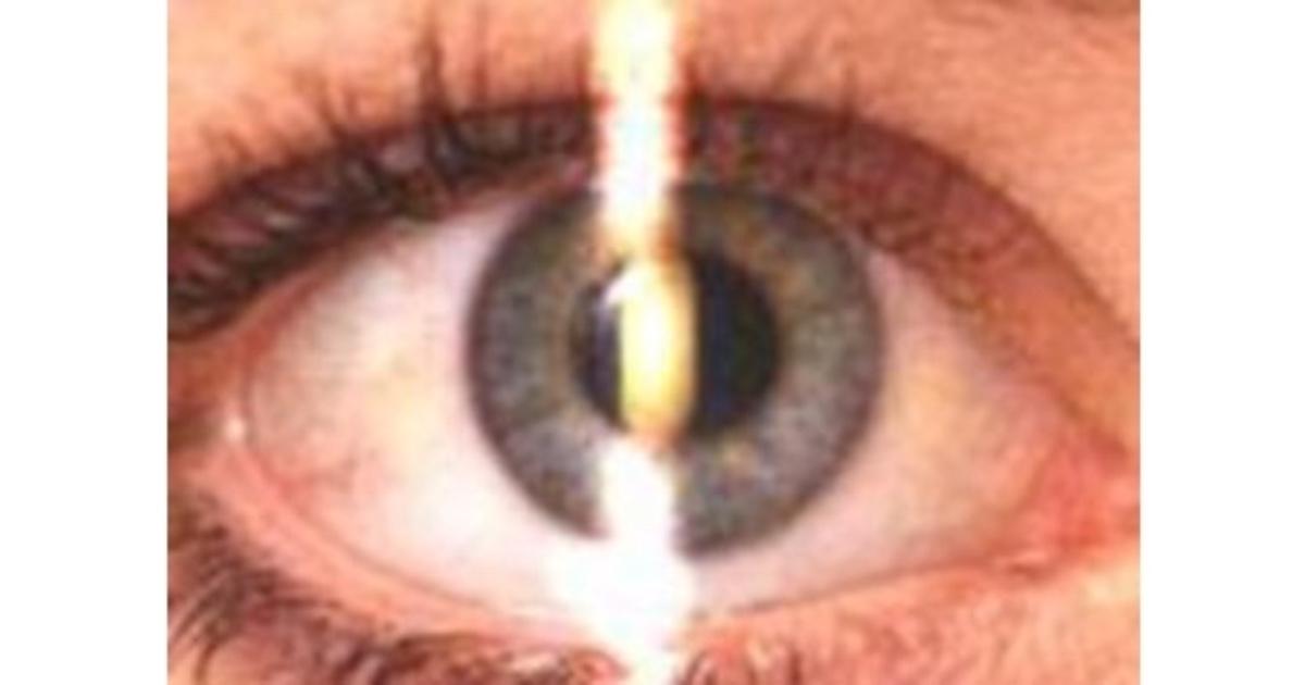
A retinoscope is a handheld medical device that eye doctors use to measure how light reflects off the retina to determine an eyeglass or contact lens prescription
What It Does
Objective testing: Measures refractive errors without requiring the patient to speak or respond.
Detects conditions: Identifies nearsightedness (myopia), farsightedness (hyperopia), and astigmatism.
Patient flexibility: Ideal for infants, young children, and individuals with communication or cognitive difficulties
How the Test Works
Shining light: The doctor projects a beam of light through your pupil onto your retina at the back of the eye.
Observing movement: They tilt the instrument back and forth to watch how the reflected red light (the retinoscopic reflex) moves.
Using lenses: The doctor places various corrective lenses in front of your eye until the reflection stops moving and fills the pupil entirely—a point known as "neutrality".
Calculating prescription: The power of the lens needed to achieve neutrality reveals your accurate prescription
Types of Retinoscopy
There are two main types of retinoscopy, static and dynamic. Static retinoscopy requires the patient have relaxed accommodation which can be achieved by focusing on a distant object or through cycloplegic agents. Meanwhile, dynamic retinoscopy requires the patient have active accommodation by focusing on a near object and is useful to evaluate the effectiveness of accommodation.
Principles of Static Retinoscopy
SpringerLink
Retinoscopy determines a patient’s refractive error by establishing what corrective lens places the far point of the eye at infinity. Infinity is simulated at the peephole of the retinoscope where all light beams reflecting from the patient’s eye are gathered. The far point is the point where light from the retinoscope focuses after reflecting off the retina. It is defined as the point in space that is conjugate with the retina in a non-accommodating eye.
When the far point is located behind the retinoscope or behind the retina, no change in the direction of light will be observed, creating a “with motion” reflex. When the far point is located between the retinoscope and the patient’s eye, this point acts as a fulcrum, creating an “against motion” reflex. When the far point is in the plane of the retinoscope, neutrality is observed with no motion of the reflex.
There exist four main refractive states of the eye: emmetropia, myopia, hyperopia, and astigmatism. Emmetropia (normal vision) is a refractive state wherein parallel light rays are focused on the fovea of a non-accommodating eye and emerge as parallel light rays. If this is the case, neutralization will be observed. Myopia consists of light rays that come to a point of focus in front of the retina in a non-accommodating eye and emerge as converging light rays. “Against motion” is observed in uncorrected myopic patients. Hyperopia consists of light rays that come to a point of focus behind the retina in a non-accommodating eye and emerge as diverging light rays. Hyperopia produces “with motion” on retinoscopy in optically uncorrected patients. Lastly, with astigmatism, light rays form two points of focus in the eye. Astigmatism can produce either a “with” and/or “against motion” reflex.
The location of the far point can be altered based on the correcting lens placed in front of the eye. Patients with emmetropia will already have the far point at infinity since the rays reflect parallel from the eye and require no correcting lens. Meanwhile, patients with myopia will have a far point between the patient’s eye and infinity since the rays leave the eye converging, requiring a minus lens to push the far point outwards. Patients with hyperopia will have a far point beyond infinity since the rays reflect diverging from the eye, requiring a plus lens to pull the focal point towards the eye.
Principles of Dynamic Retinoscopy
Dynamic retinoscopy permits a rapid, objective assessment of a patient’s accommodative ability. Unlike static retinoscopy, accommodation is not relaxed when assessing dynamic retinoscopy nor are lenses used. The goal of this technique is to encourage the patient to use their accommodation to focus on a target in line with the peep hole of the retinoscope.
Dynamic Retinoscopy Technique
Set up
Seat the patient in the examination chair, in an upright position. The patient should be wearing their appropriate distance optical correction.
Dim the room lights enough to better see the retinoscopic reflex but to still allow the patient to see the near target. A dim reading light can be used to facilitate viewing of the near target.
Hold the retinoscope at a reading distance from the patient.
Hold a near target (small letters or a sticker with small details) in line with the peep hole of the retinoscope.
Illustration of the position of the patient’s fixation, retinoscope, and associated light rays.
Examination technique
Ask the patient to fixate on a distance target.
Sweep the retinoscope at both 90 and 180 degrees and observe the patient’s retinoscopic reflex. Move back and forth between both eyes. “With” motion should be observed in both eyes. Check both meridians for presence of astigmatism.
Ask the patient to then fixate on the near target (held at approximately reading distance).
Repeat the sweep of the retinoscope at both 90 and 180 degrees and observe the patient’s retinoscopic reflex. Move back and forth between both eyes. The previous “with” motion reflex should rapidly change to a slight “against” motion reflex. If this is not observed, encourage the patient to focus on a smaller part of the target or move back slightly yourself.
Repeat steps 2-4 to ensure accommodation is sustainable for longer periods.