Dechrome test
Introduction
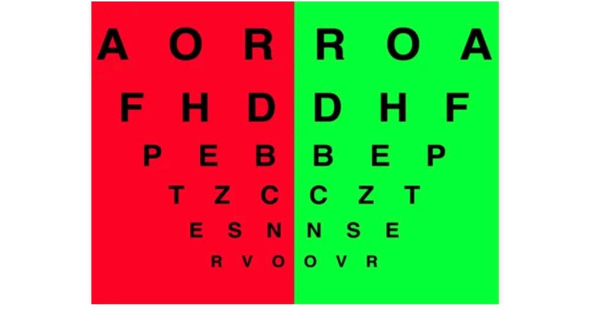
Chromatic aberration, the basis of the test, occurs because different wavelengths of light are bent to a different extent. The duochrome test involves the projection of black letters or symbols onto a bipartite green (at approximately 535nm) and red (at approximately 620 nm) background. The red and green wavelengths are dioptrically equidistant, approximately 0.25 D, from the yellow wavelength (570 nm). The longer wavelength (red) is refracted less than the shorter (green). It is assumed that best vision is attained when the yellow wavelengths are focused on the retina. During the final sphere adjustment, it is important to find the least minus that a patient will accept in order to ensure that accommodation is minimized, particularly in myopic patients. The eye typically focuses near the midpoint of the spectrum, between the red and green wavelengths. With optimal spherical correction, the letters on the red and green halves of the chart appear equally sharp.
Indications
Contraindications
The duochrome test is not used with patients whose visual acuity is worse than 20/40, because the 0.50 D difference between the 2 sides is too small to distinguish.
Equipment
Procedure
The patient is first assessed with refraction and given the best correction.
With that correction in the phoropter or trial frames in place, the duochrome chart can then be projected.
The test starts by occluding one eye (left eye first by convention)
Room lights must be turned off in order to allow the pupil to dilate which would allow for increase chromatic aberration of the eye
Ask the patient to compare the letters on green and red sides of the test
If the letters on the red side stand out more continue to add more minus power using -0.25 DS steps
If the letters on the green side stand out more continue to add more plus power using +0.25 DS steps
Neutrality is reached when the letters on both the red and the green background appear equally distinct.
IF more than +/- 0.5D is required to balance then the test is unreliable
The RAM-GAP Rule
Doctors use a mnemonic to interpret the patient's response: RAM-GAP.
RAM (Red Adds Minus) - If the patient says the letters on the Red side are sharper, it means the eye is slightly myopic (nearsighted) with the current lens. The image focus is in front of the retina. The doctor adds more Minus power to push the focus back.
GAP (Green Adds Plus) - If the patient says the letters on the Green side are sharper, it means the eye is slightly hyperopic (farsighted) or over-minused. The image focus is behind the retina. The doctor adds Plus power (or reduces minus) to pull the focus forward.